Human Muscle
Muscle is made up of thousands of muscle fibers, each composed of a single muscle cell.
As shown in the Figure below a muscle cell contains a series of ultramicroscopic filaments called myofibrils.
Each myofibril is a muscle cell that contains units called sarcomeres.
Sarcomeres contain thick microfilaments composed of the protein myosin.
Sarcomeres also contain thin microfilaments composed of the protein actin.
The actin and myosin filaments are arranged parallel to one another, with the myosin filaments’ molecular “heads” protruding toward the actin filaments.
In skeletal muscle, the overlapping actin and myosin filaments give the muscle fiber a banded, or striated, appearance.
Hence, the muscle is striated muscle.
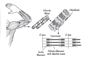
Figure above shows The Anatomical structure of the muscle.
Muscle contraction
When a nerve impulse arrives at the muscle cells, it passes across the neuromuscular junction and enters the muscle cell membrane, which is known as the sarcolemma.
The impulse spreads across the muscle cell and enters its cytoplasm, which is called sarcoplasm.
The nerve impulse causes the actin filaments to slide across the surface of the myosin filaments.
The sliding filaments pull together the ends of the muscle cell, thereby causing it to contract.
The sliding filaments require that calcium ions and energy in the form of ATP be available.
Two proteins called tropomyosin and troponin also function in the contraction.
Cross-bridges hold the filaments together as the muscle contracts.
After the muscle contraction has taken place, the energy to sustain the contraction is used up, and the cross-bridges break.
The filaments then slide back to their original position, and the muscle cell relaxes.
There is no partial contraction of the muscle cell.
Contraction is an all-or-none phenomenon.
Energy for contraction
Adenosine triphosphate (ATP) supplies the energy for muscle contraction.
The reactions of glycolysis, the Krebs cycle, and the electron transport system normally produce ATP during cellular respiration.
During normal activity, ATP is regenerated as it is used up during muscle contraction.
When a
Creatin phosphate transfers its energy to new ATP molecules, which then function as additional energy sources.
When creatin phosphate is used up, muscle cells obtain their energy solely from the process of glycolysis.
Because no oxygen is available, the metabolism is anaerobic.
Under these conditions, two molecules of ATP are obtained per molecule of glucose metabolized.
The pyruvic acid that forms is converted to lactic acid in the muscle.
Lactic acid prevents overexertion of the muscle because as lactic acid accumulates, the person experiences fatigue.
The fatigue induces the person to stop exerting the muscles and breathe deeply.
This breathing provides a plentiful supply of oxygen to satisfy the oxygen debt.
Lactic acid is converted back to pyruvic acid, which is then metabolized through the Krebs cycle and electron transport system to provide a new supply of ATP and creatin phosphate.
Types of muscle
The human body has three major types of muscle.
The muscle type discussed earlier in this chapter is striated muscle because the fibers’ overlapping actin and myosin filaments give it a banded appearance.
This muscle is found in the limbs and is also called skeletal muscle.
It operates under voluntary control and so is additionally known as voluntary muscle.
The second muscle type is smooth muscle, which has few actin and myosin filaments; therefore, it has few striations.
Smooth muscle is found in the linings of the blood vessels, along the gastrointestinal tract, in the respiratory tract, and in the urinary bladder.
Because it operates without voluntary control, it is sometimes called involuntary muscle.
The third muscle type is cardiac muscle, which is found in the heart.
It has striations because it has multiple actin and myosin filaments, but it is an involuntary muscle.
The actin and myosin filaments in cardiac muscle exist as intertwined branches that form a conducting network for nerve impulses.
Army Jobs - Navy Jobs - Military Jobs

Scholarship 2026/27
Current Scholarships 2026/2027 - Fully Funded
Full Undergraduate Scholarships 2026/2027
Fully Funded Masters Scholarships 2026/27
PhD Scholarships for International Students - Fully Funded!
Funding Opportunities for Journalists 2026/2027
Funding for Entrepreneurs 2026/2027
Scholarship 2026/27
Current Scholarships 2026/2027 - Fully Funded
Full Undergraduate Scholarships 2026 - 2027
Fully Funded Masters Scholarships 2026 - 27
PhD Scholarships for International Students - Fully Funded!
Funding Opportunities for Journalists 2026/2027
Funding for Entrepreneurs 2026/2027
***