Cell Cycle
The cell cycle involves many repetitions of cellular growth and reproduction.
With few exceptions (for example, red blood cells), all the cells of living things undergo a cell cycle.
The cell cycle is generally divided into two phases: interphase and mitosis.
During interphase, the cell spends most of its time performing the functions that make it unique.
Mitosis is the phase of the cell cycle during which the cell divides into two daughter cells.
Interphase
The interphase stage of the cell cycle includes three distinctive parts: the G1 phase, the S phase, and the G2 phase.
The G1 phase follows mitosis and is the period in which the cell is synthesizing its structural proteins and enzymes to perform its functions.
For example, a pancreas cell in the G1 phase will produce and secrete insulin, a muscle cell will undergo the contractions that permit movement, and a salivary gland cell will secrete salivary enzymes to assist digestion.
During the G1 phase, each chromosome consists of a single molecule of DNA and its associated histone protein.
In normal human cells, there are 46 chromosomes per cell (except in sex cells with 23 chromosomes and red blood cells with no nucleus and, hence, no chromosomes).
During the S phase of the cell cycle, the DNA within the nucleus replicates.
During this process, each chromosome is faithfully copied, so by the end of the S phase, two DNA molecules exist for each one formerly present in the G1 phase.
Human cells contain 92 chromosomes per cell in the S phase.
In the G2 phase, the cell prepares for mitosis.
Proteins organize themselves to form a series of fibers called the spindle, which is involved in chromosome movement during mitosis.
The spindle is constructed from amino acids for each mitosis, and then taken apart at the conclusion of the process.
Spindle fibers are composed of microtubules.
Mitosis
The term mitosis is derived from the Latin stem mito, meaning “threads.
” When mitosis was first described a century ago, scientists had seen “threads” within cells, so they gave the name “mitosis” to the process of “thread movement.”
During mitosis, the nuclear material becomes visible as threadlike chromosomes.
The chromosomes organize in the center of the cell, and then they separate, and 46 chromosomes move into each new cell that forms.
Mitosis is a continuous process, but for convenience in denoting which portion of the process is taking place, scientists divide mitosis into a series of phases: prophase, metaphase, anaphase, and telophase (see the figure below):
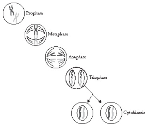
The process of mitosis, in which the chromosomes of a cell duplicate and pass into two daughter cells.
Prophase: Mitosis begins with the condensing of the chromatin to form chromosomes in the phase called prophase.
. Two copies of each chromosome exist; each one is a chromatid.
Two chromatids are joined to one another at a region called the centromere.
As prophase unfolds, the chromatids become visible in pairs (called sister chromatids), the spindle fibers form, the nucleoli disappear, and the nuclear envelope dissolves.
In animal cells during prophase, microscopic bodies called centrioles begin to migrate to opposite sides of the cell.
When the centrioles reach the poles of the cell, they produce and are then surrounded by a series of radiating microtubules called an aster.
Centrioles and asters are not present in most plant or fungal cells.
As prophase continues, the chromatids attach to spindle fibers that extend out from opposite poles of the cell.
The spindle fibers attach at the region of the centromere at a structure called the kinetochore, an area of protein in the centromere region.
Eventually, all pairs of chromatids reach the center of the cell, a region called the equatorial plate.
Metaphase: Metaphase is the stage of mitosis in which the pairs of chromatids line up on the equatorial plate.
This region is also called the metaphase plate.
In a human cell, 92 chromosomes in 46 pairs
Each pair is connected at the centromere, where the spindle fiber is attached (more specifically at the kinetochore).
Anaphase: At the beginning of anaphase, the sister chromatids move apart from one another.
The chromatids are called chromosomes after the separation.
Each chromosome is attached to a spindle fiber, and the members of each chromosome pair are drawn to opposite poles of the cell by the spindle fibers.
During anaphase, the chromosomes can be seen moving.
They take on a rough V shape because of their midregion attachment to the spindle fibers.
The movement toward the poles is accomplished by several mechanisms, such as an elongation of the spindle fibers, which results in pushing the poles apart.
The result of anaphase is an equal separation and distribution of the chromosomes.
In human cells, a total of 46 chromosomes move to each pole as the process of mitosis continues.
Telophase: In telophase, the chromosomes finally arrive at the opposite poles of the cell.
The distinct chromosomes begin to fade from sight as masses of chromatin are formed again.
The events of telophase are essentially the reverse of those in prophase.
The spindle is dismantled and its amino acids are recycled, the nucleoli reappear, and the nuclear envelope is reformed.
Cytokinesis: Cytokinesis is the process in which the cytoplasm divides and two separate cells form.
Note that cytokinesis is separate from the four stages of mitosis.
In animal cells, cytokinesis begins with the formation of a cleavage furrow in the center of the cell.
With the formation of the furrow, the cell membrane begins to pinch into the cytoplasm, and the formation of two cells begins.
This process is often referred to as cell cleavage.
Microfilaments contract during cleavage and assist the division of the cell into two daughter cells.
In plant cells, cytokinesis occurs by a different process because a rigid cell wall is involved.
Cleavage does not take place in plant cells.
Rather, a new cell wall is assembled at the center of the cell, beginning with vesicles formed from the Golgi apparatus (see Chapter 3).
As the vesicles join, they form a double membrane called the cell plate.
The cell plate forms in the middle of the cytoplasm and grows outward to fuse with the cell membrane.
The cell plate separates the two daughter cells.
As cell wall material is laid down, the two cells move apart from one another to yield two new daughter cells.
Mitosis serves several functions in living cells.
In many simple organisms, it is the method for asexual reproduction (for example, in the cells of a fungus).
In multicellular organisms, mitosis allows the entire organism to grow by forming new cells and replacing older cells.
In certain species, mitosis is used to heal wounds or regenerate body parts.
It is the universal process for cell division in eukaryotic cells.
Army Jobs - Navy Jobs - Military Jobs

Scholarship 2026/27
Current Scholarships 2026/2027 - Fully Funded
Full Undergraduate Scholarships 2026/2027
Fully Funded Masters Scholarships 2026/27
PhD Scholarships for International Students - Fully Funded!
Funding Opportunities for Journalists 2026/2027
Funding for Entrepreneurs 2026/2027
Scholarship 2026/27
Current Scholarships 2026/2027 - Fully Funded
Full Undergraduate Scholarships 2026 - 2027
Fully Funded Masters Scholarships 2026 - 27
PhD Scholarships for International Students - Fully Funded!
Funding Opportunities for Journalists 2026/2027
Funding for Entrepreneurs 2026/2027
***